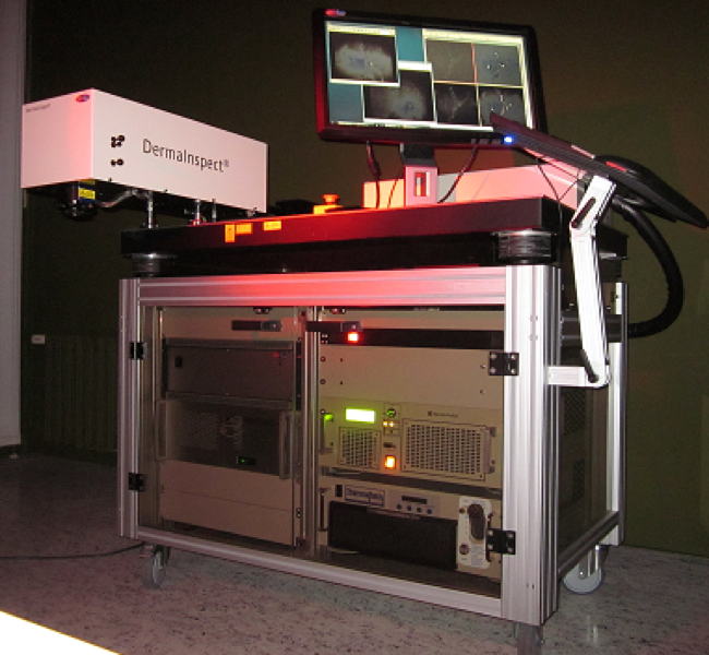
The historical multiphoton tomograph DermaInspect® is an imaging device that provides non-invasive in vivo optical biopsies of skin with ultrahigh subcellular resolution. The 1M class system uses a femtosecond laser beam for multiphoton excitation of biomolecules like NAD(P)H, flavins, porphyrins, elastin, and melanin. The extracellular matrix element collagen is imaged by its second harmonic generation (SHG). Autofluorescence and SHG signals are recorded by fast PMT detectors.
The system consists of a compact, turn-key tunable femtosecond near-infrared (NIR) laser, a beam scanning module with galvoscanners and piezo-driven optics, a PMT detector module as well as a control unit including JenLab Image software for 3D, 4D and 5D image processing.
FLIM Technology
In addition to 3D fluorescence imaging by optical sectioning and monitoring of the fluorescence intensity, DermaInspect® system can be upgraded with a FLIM module of an external manufacturer for fluorescence lifetime imaging. DermaInspect® combined with the time-correlated single photon counting (TCSPC) module can perform fluorescence lifetime imaging (FLIM) at different tissue depths with a temporal resolution of 250 ps. The fluorescence lifetime τ adds a 4th dimension to the high-resolution images and provides information on the type of fluorescent biomolecule as well as molecular
interactions within the microenvironment. For example, typical fluorescence lifetimes of free NADH, NADH-protein complexes and porphyrin monomers are 200 ps, 2 ns, and 10 ns, respectively.
Spectral Imaging
The multiphoton tomograph DermaInspect® can be further upgraded to a 5D system in order to obtain information on the emission spectrum (spectral imaging with submicron spatial resolution).
A CE-marked system for in vivo multiphoton tomography of skin with submicron spatial resolution
In vivo optical biopsies with subcellular spatial resolution based on near infrared femtosecond laser technology for:
- diagnostics of dermatological disorders
- melanoma detectiont
- tissue engineering
- cosmetic research
- in situ drug monitoring
- intratissue imaging of pharmaceutical components
Applications
The major aim for the development of the DermaInspect® was skin cancer diagnosis. Using innovative non-invasive multiphoton technology the physician obtains detailed information on the living intratissue cells and the tissue architecture within their natural environment. Dermatological disorders and melanoma can now be detected with submicron spatial resolution. Having a short acquisition time of seconds, DermaInspect® revolutionizes conventional invasive and highly time consuming diagnostic procedures. The system is used in tissue engineering for the detection of cells and extracellular matrix components, in the research of wound healing and skin aging, in monitoring of therapeutical effects as well as in cosmetic and pharmaceutical research. FLIM and spectral imaging provide further contrast mechanisms for in situ drug monitoring. Researchers using this combination will have an exclusive fourdimensional view on biological processes within living tissue.
DermaInspect® is routinely used in various research institutes and hospitals in Europe, Asia and Australia.
References
K. König, I. Riemann: „High-resolution multiphoton tomography of human skin with sub cellular spatial resolution and picosecond time resolution.” Journal of Biomedical Optics 8, 432-439 (2003)
K. König, A. Ehlers, F. Stracke, I. Riemann: „In vivo Drug Screening in Human Skin Using Femtosecond Laser Multiphoton Tomography.” Skin Pharmacology and Physiology 19, 78-88 (2006)
K. König, I. Riemann, A. Ehlers, R. Bückle, E. Dimitrow, M. Kaatz, J. Fluhr, P. Elsner: „In vivo multiphoton tomography of skin cancer.” Proc. of SPIE Vol. 6089, pp 118-124 (2006)
M.J. Koehler, K. König, P. Elsner, R. Bückle, M. Kaatz: „In vivo assessment of human skin aging by multiphoton laser scanning thomography.“ Optics Letters 31, 2879-2881 (2006)
Technical Data
- compact turn-key tunable Ti:sapphire femtosecond laser
- laser pulse width: < 100 fs
- repetition frequency: 80 / 90 MHz
- mean laser output: 0 ... 1.5 W
- wavelength range: 720 ... 920 nm
- full-frame scanning, region-of-interest (ROI) line scanning, single-point illumination (spot scan)
- typical scan range: 350 × 350 μm (horizontal), 200 μm (vertical)
- spatial resolution: < 1 μm (horizontal), < 2 μm (vertical)
- temporal resolution (FLIM): 250 ps
- focusing optics: magnification 40×, numerical aperture (NA) 1.3
- video adapter for visualization with CCD-camera
- control and image processing software (JenLab Image)
- operating temperature: 15 ... 35 °C (59 ... 95 °F)
- relative humidity: 5 ... 65 % (non-condensing)
- power requirements: 230 VAC (50 Hz) or 115 VAC (60 Hz)
- CE certified
System dimensions
- Workstation: 1200 × 780 × 1100 mm3, 180 kg
- Scan module: 615 × 255 × 210 mm3, 16 kg
- Control unit: 450 × 460 × 190 mm3, 12 kg
- Ti:sapph. laser: 600 × 370 × 180 mm3, 42 kg (laser head), 41 kg (power supply)
- Chiller: 270 × 200 × 380 mm3, 20 kg
The complete system needs a minimum space of 8 m². Air-conditioning is recommended. System operation requires reduction of ambient light.
Notes: These specifications are subject to change without notice.
DermaInspect® is not intended as a primary diagnostic means.
PDF-Download: Summary of the mirror arm